Keep Your Brain 11 Years Younger With Leafy Green Vegetables
While cognitive abilities naturally decline with age, eating one serving of leafy green vegetables a day may aid in preserving memory and thinking skills as a person grows older, according to a study by researchers at Rush University Medical Center in Chicago. The study results were published in the December 20, issue of Neurology, the medical journal of the American Academy of Neurology.
“Adding a daily serving of green leafy vegetables to your diet may be a simple way to help promote brain health,” said study author Martha Clare Morris, ScD, a nutritional epidemiologist at Rush. “There continues to be sharp increases in the percentage of people with dementia as the oldest age groups continue to grow in number. Effective strategies to prevent dementia are critically needed.”
The study results suggest that people who ate one serving of green, leafy vegetables had a slower rate of decline on tests of memory and thinking skills than people who rarely or never ate them. The study results also suggest that older adults who ate at least one serving of leafy green vegetables showed an equivalent of being 11 years younger cognitively.
960 older adults completed food questionnaires and received annual cognitive assessments
The study enlisted volunteers already participating in the ongoing Rush Memory and Aging Project, which began in 1997 among residents of Chicago-area retirement communities and senior public housing complexes. A “food frequency questionnaire” was added from 2004 to February 2013, which 1,068 participants completed. Of them, 960 also received at least two cognitive assessments for the analyses of cognitive change.
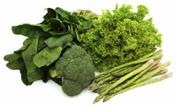
This study involved these 960 people, who at the study start were an average age of 81 years old and did not have dementia. They had their thinking and memory skills tested every year and were followed for an average of 4.7 years. The participants also completed the food frequency questionnaire, which assessed how often and how many half-cup servings they ate of either spinach; kale/collards/greens; or a one-cup serving of lettuce/salad.
The study divided the participants into five groups based on how often they ate green leafy vegetables, and compared the cognitive assessments of those who ate the most (an average of about 1.3 servings per day) and those who ate the least (0.1 servings per day).
Overall, the participants’ scores on the thinking and memory tests declined at a rate of 0.08 standardized units per year. Over 10 years of follow-up, the rate of decline for those who ate the most leafy greens was slower by 0.05 standardized units per year than the rate for those who ate the least leafy greens. This difference was equivalent to being 11 years younger in age, according to Morris.
More research needed in younger and minority populations
The results remained valid after accounting for other factors that could affect brain health, such as seafood and alcohol consumption, smoking, high blood pressure, obesity, education level and amount of physical and cognitive activities.