Mature Cells Turned Into Stem Cells Without Genetic Engineering
Research by the National University of Singapore and the FIRC Institute of Molecular Oncology in Italy has revealed that mature cells can be reprogrammed into redeployable stem cells without genetic engineering. This is important because there are significant health risks associated with treatment utilizing stem cells with added genes. Instead of direct genetic modification, they are instead confined to a defined geometric space for an extended period of time. The breakthrough findings will no doubt usher in a new generation of technologies for stem cell tissue engineering and regenerative medicine.
In earlier studies over a decade ago scientists showed that mature cells could be reprogrammed to become pluripotent stem cells which were capable of being developed into any cell type in the body. Scientists accomplished this by genetically modifying mature cells by introducing external factors that would reset genomic programs of the cells. This essentially turned back the clock returning them to an undifferentiated or unspecified state. These lab made cells which are known as induced pluripotent stem cells (iPSCs) could then be programmed into different types of cells for tissue repair, drug discovery and also to grow new organs for transplants. These cells did not need to be harvested from embryos.
Unfortunately a major problem is the tendency for any specialized cell that has been developed from iPSCs to form tumors after introduction into the body. The researchers turned their focus to understanding how stem cell growth and differentiation is regulated in the body and how cells naturally convert to another cell type or revert to an immature stem cell like state during tissue maintenance or during development.
The new research has shown that mature cells can be reprogrammed in vitro into pluripotent stem cells by confining the cells to a defined area for growth rather than generically modifying them.
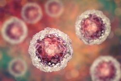
When fibroblast cells which are a type of mature cell found in connective tissue such as ligaments and tendons were confined to rectangular areas, they very quickly assumed the shape of the the surface of the medium that they were attached to. This indicated the cells were responding and measuring to the physical properties of the environment then conveying this information to the nucleus where DNA genome programs and packaging would adapt accordingly.
The cells were grown over ten days until they formed spherical clusters of cells. The genetic analysis of the cells contained within these clusters revealed that specific characteristics of chromatin which is the condensed from of packaged DNA normally associated with mature fibroblasts were lost by the 6th day. At the 10th day the cells expressed genes which are normally associated with embryonic stem cells and iPSCs. They team learned that by confining the mature cells for an extended period the mature fibroblasts could be turned into pluripotent stem cells. To confirm, the researchers directed their growth with high efficiency into two different specialized cell types and some cells were also directed back into fibroblasts.
The physical parameters used are reflective of the transient geometric constraints that cells can be exposed to in the body. During development the establishment of niches and geometric patterns are essential to the formation of functional organs and tissues. When tissue is damaged through disease or injury, cells will experience sudden alterations to their environment. In these cases, mature cells might revert back to a pluripotent stem cell like state prior to being redeployed as specialized cells for maintenance and repair of tissue. The study shows for the first time that mechanical cues can reset genomic programs of mature cells and then return them to a pluripotent state.
The researchers believe the use of geometric constraints to reprogram mature cells might better reflect processes that naturally occur within the body. And more importantly, the findings allow researchers to generate stem cells from mature cells with high efficiency and without genetically modifying them.
To view the original scientific study click here: Laterally confined growth of cells induces nuclear reprogramming in the absence of exogenous biochemical factors