Breakthrough in Genetic Editing

Gene Editing

Imagine being able to have your genes edited to eradicate errors and to greatly lengthen quality of life and life span. There are some people that are still active and independent at the age of 100 and live well beyond that. Generally those people also had long lived parents. What if the same genetic traits they inherited and more could be given to anyone?

Researchers at the Joint Institute of Metrology and Biology (JIMB) have recently developed MAGESTIC, a new CRISPR platform that is similar to a word processor which makes gene editing using barcodes. CRISPR which snips at DNA has been a clumsy tool making precision medicine or ?clipping? disease causing genetic mutations in patients virtually impossible. MAGESTIC or ?multiplexed accurate genome editing with short, trackable, integrated cellular barcodes? is being compared to a word processer that enables efficient search and replace functions for genetic material. This new platform was also able to produce an increase of sevenfold in survival of cells during the process of editing.

Previously, CRISPR required a very extensive understanding of how repair cuts with cells at a variety of sites across the genome could be controlled as needed. Because DNA strands are able to rejoin in very unpredictable ways, random mutations are likely to occur at the cut sites in the DNA of the cells. Additionally, many cells will not survive the process of editing at all. It has been extremely challenging building very accurate predictions of gene editing. Researchers want a more reliable way for programming CRISPR to be able to cut at targeted locations in the genome and then be able to direct cells for designed edits at the cut sites of the DNA. This can be accomplished by providing a donor DNA for the cell which the cell?s DNA repair machinery is able to use as a template for replacing the original sequence at the original cut site. However, the cell?s DNA repair system is complex and will not always behave predictably.

The cell searching for a DNA donor suitable for repairing a cut site is a huge challenge for the cell. The repair machinery of the DNA has to search through millions and even billions of DNA base pair sequences to be able to find the correct donor DNA. MAGESTIC has provided a significant advance in gene-editing technology by helping the cell search by artificially recruiting the DNA designed donor through a process called active donor recruitment which will recruit the donor DNA right to the cut site. This recruitment resulted in a sevenfold increase in the cell?s survival which was a change that resulted in increased effectiveness and efficiency.

The other feature that was different from CRISPR, was MAGESTIC?S new version of cellular barcode. Previously, researchers used small bits of circular DNA also known as plasmids to guide DNAs and to store barcodes for tracking designed mutations to each cell. The plasmids will multiply with cell growth and are inherited by both cells following cell division. With MAGESTIC the barcodes are integrated into chromosomes as opposed to single barcode per item correspondence which can vary widely in number resulting in 10 to 40 appearing in every cell.

Scientists do not know much about the function of the 0.l% of code that will vary between individuals in any population and is responsible for differences in susceptibility of disease. MAGESTIC helps to address the gap in understanding natural genetic variation through enabling individual genetic variant to be edited very precisely and compared to other genetic variants one by one. This results in help in uncovering which genetic differences will have cellular impact function. MAGESTIC will also edit all at one time in just a single test tube with every edit happening in any one of a million otherwise cells that are identical.

The researchers have reached a state where they have achieved sequencing the order of genome base pairs and are also able to change them. Additional research is needed to understand the edit sequencing.

To view the original scientific study click here: Multiplexed precision genome editing with trackable genomic barcodes in yeast

Fasting Boosts Gut Health

Fasting

A 24 hour fast can reverse age related decline of stem cell function in the gut according to a study conducted by biologists at the Massachusetts Institute of Technology. The study reveals that a metabolic switch occurs during a 24 hour fast. This switch results in a break down by cells of fatty acids rather than glucose. This change boosts gut stem cell regeneration in both old and young mice.

With age stem cells in the intestine start to lose their regeneration ability. This reduction makes it more difficult for people to recover from gastrointestinal conditions and infections since the stem cells in the intestine are the source for new cells. The biologists discovered they could boost stem cell regeneration using a molecule which activates the identical metabolic switch in the stem cells to induce burning fat rather than utilizing carbohydrates. This intervention could possibly be used for people who are recovering from a variety of conditions that effect the GI tract.

Fasting provides many positive effects in the intestines. It boosts regeneration of stem cells and can be useful in a variety of ailments which affect the intestines. Switching the intestinal stem cells to fatty acid oxidation significantly enhanced their function.

Omer H. Yilmaz, Assistant Professor of Biology at MIT, refers to stem cells in the intestinal wall as ?workhorses of the intestines?. And diet has a profound effect on the regeneration ability of tissue. It is known that a low calorie diet enhances longevity in humans. Intestinal stem cell function declines with aging which therefore interferes with the intestines ability to repair after damage. The researchers decided to focus on how a 24 hour fast might enhance the function of both old and young intestinal stem cells.

Once mice had fasted for 24 hours, the team removed stem cells from the intestine and transferred them to a culture dish. Their goal was to see if the cells would rise to
?mini intestines? which are known as organoids. Interestingly the team discovered that stem cells from the fasting mice resulted in double the regenerative capacity. Fasting had a profound effect on the intestinal crypts to make more organoids which are driven by stem cells. They observed this in both aged and young mice.

Additional studies which included sequencing RNA messenger of stem cells from the fasting mice showed fasting induced cells to change from the usual metabolism of carbohydrate burning to fatty acid metabolizing. The switch that occurs is through activation of transcription factors also referred to as PPARs. These turn on genes which are part of the metabolizing of fatty acids. The team discovered that by turning off this pathway, the fasting period would no longer boost regeneration. The team was also able to reproduce the positive effects of fasting to mimic the effects of PPARs by treating the mice with a molecule which mimics these effects. By activating just one metabolic pathway they were able to reverse some age phenotypes.

The next step is to study the process of the metabolic switch which provokes stem cells to strengthen their ability to regenerate. The team also plans to further research into how fasting might affect the ability of stem cells to regenerate other tissue types.

It may be possible to stimulate regeneration without having to fast for 24 hours which can be difficult for some people. Intermittent fasting involves eating during an 8 hour period each day and then fasting for 16 hours while drinking plenty of water. Some people do this every day. For those who have trouble going without food it can help a to eat a very low sugar diet including avoidance of high sugar fruits such as bananas, dates and dried fruit. All sweet foods and sweeteners tend to stimulate appetite and cause cravings.

To view the original scientific study click here: Fasting Activates Fatty Acid Oxidation to Enhance Intestinal Stem Cell Function during Homeostasis and Aging

Zero Calorie Sweeteners Cause Weight Gain

Zero Calorie Sweeteners

In addition to common artificial sweeteners shown to change how the body processes fat and energy, a new study has revealed they can also cause health changes that are linked to obesity and diabetes. Because of the increased awareness of the health consequences associated with eating too much sugar, a dramatic increase in the consumption of zero calorie artificial sweeteners has occurred.

Artificial sweeteners are one of the most common food additives and are frequently consumed in diet and zero calorie beverages and other food products. The recent study is the largest examination to date which tracks biochemical changes in the body. Previous studies linked artificial sweeteners with negative health consequences; however the research had been mixed and raised questions about potential bias related to study sponsorship. The new study used an approach known as unbiased high throughout metabolomics after consumption of sugar and sugar substitutes. The tests were conducted in rats and cell cultures, and the researchers also looked at impacts on vascular health by studying how the substances affect the lining of blood vessels.

The research included feeding different groups of rats diets high in glucose or fructose, or aspartame or acesulfame (zero calorie artificial sweeteners). At the end of three weeks the researchers saw significant differences in the concentrations of fat, amino acids and biochemicals in blood samples. The results suggest that artificial sweeteners change how the body processes fat and gets its energy. They also found that acesulfame potassium appeared to accumulate in the blood with higher concentrations having a more harmful effect on the cells that line the blood vessels.

The researchers noted that in moderation the body can handle sugar. But when the system is overloaded over a long period the body machinery breaks down. They observed that replacing those sugars with the zero caloric artificial sweeteners led to negative changes in energy and fat metabolism.

The researchers note that the results do not provide a clear answer and further study is needed. But the question becomes, which is worse between sugar or artificial sweeteners? It is well known that high dietary consumption of sugar is related to negative health outcomes. The recent study suggests that the artificial sweeteners do too. If people chronically consume the foreign substances as with sugar also, the risk of health problems such as obesity and diabetes increases.

Experimental Biology 2018. “Why zero-calorie sweeteners can still lead to diabetes, obesity: Common artificial sweeteners shown to change how the body processes fat and energy.” ScienceDaily. ScienceDaily, 23 April 2018.

Raw Fruits and Vegetables are Good for Your Brain

raw-fruits-vegetables
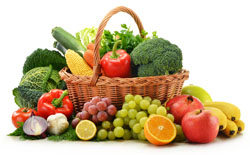
A new study conducted by the University of Otago has revealed that fruits and vegetables consumed in their natural state promote better?brain health. The study which was published in Frontiers in Psychology found that for mental health in particular these food items in their ?unmodified? state retained more of their nutrients as opposed to consuming them cooked or canned.

A total of 400 young adults aged 18 to 25 from the United States and New Zealand participated in the study. This age group was chosen as they typically have the lowest vegetable and fruit consumption of all age groups and are additionally are at high risk for mental health disorders. The individual?s typical consumption of raw fruits and vegetables vs. cooked or canned were assessed. Their positive and negative mental health, lifestyle and demographic variables that could affect the association between vegetable and fruit intake were also taken into consideration such as health styles including sleep, exercise, unhealthy diet, chronic health conditions, ethnicity, gender and socioeconomic status.

Raw fruit and vegetable vs. canned or cooked fruits and vegetables predicted lower levels of mental illness symptomology such as depression, and improved levels of psychological wellbeing such as positive mood, life flourishing and satisfaction. The researchers found that these mental health benefits were significantly reduced for the canned and cooked fruits and vegetables indicating that consuming them in their natural state was more beneficial.

The top 10 raw foods related to better health were bananas, apples, carrots, dark leafy greens, grapefruit, citrus fruits, lettuce, fresh berries, kiwi fruit and cucumber. The research is increasingly beneficial as dietary changes may provide a safe, accessible and adjuvant approach to improving mental health.

An earlier study conducted by the University of Warwick?s Medical School which stressed the importance of consuming 5 or more fruits and vegetables per day, found that 33.5% of participants with high mental health wellbeing consumed at least that many servings per day. This compared to only 6.8% who ate less than one portion per day. This study did not focus on how the fruits and vegetables were consumed (fresh vs. cooked or canned), but additionally supports the findings of the importance of consuming 5 or more portions per day of these nutrient dense foods. This study found that along with smoking, fruit and vegetable consumption was the health-related behavior most consistently associated with high and low mental wellbeing. Both studies show that consuming fruits and vegetables may plan a potential role as a driver of not only better physical health, but also of mental wellbeing.

References:

Kate L. Brookie, Georgia I. Best, Tamlin S. Conner. Intake of Raw Fruits and Vegetables Is Associated With Better Mental Health Than Intake of Processed Fruits and Vegetables. Frontiers in Psychology, 2018; 9 DOI: 10.3389/fpsyg.2018.00487

S. Stranges, P. C. Samaraweera, F. Taggart, N.-B. Kandala, S. Stewart-Brown. Major health-related behaviours and mental well-being in the general population: the Health Survey for England. BMJ Open, 2014; 4 (9): e005878 DOI: 10.1136/bmjopen-2014-005878

New Stem Cell Niche Discovery

Adult Stem Cells

Researchers at Duke NUS Medical School headed by Gedas Greicius and Professor David Virshup, Director of the Programme in Cancer and Stem Cell Biology, have identified how the stem cell neighborhood, or niche, keep stem cells alive in the gut.

Stem cells have the ability to differentiate or develop into many different cell types in the body. They also serve as a repair system to replace damaged or aged cells. With the ability to regenerate, stem cells offer enormous potential to improve health, quality of life and lifespan.

Stem cells in our adult tissues live in very specific locations called stem cell niches where they provide an ideal and specialized neighborhood for stem cells. The stem cells in the niche are undifferentiated which means they have not changed into mature cells. This stem cell niche regulates how stem cells participate in tissue maintenance, regeneration and repair. The niche prevents stem cells from being depleted while also protecting the body from an over production of the stem cells. This understanding of stem cell niches is important in the field of stem cell therapeutics.

To understand the role of the niche, the researchers needed to identify the key cell types that regulate the numerous processes that take place within the cell niche. The key regulators in the intestinal cell niche are hormones called R-spondins and Wnts which are frequently expressed together. It has been unclear what type of niche cells make the R-spondins and Wnts.

The team studied the source and functional role of RSPO3, a R-spondin, and Wnts. RSPO3 is by far the most abundant R-spondin produced in the small intestine of a mouse. Using a mouse model, they identified the specific cell called a subepithelial myofibroblast as an essential source of both RSPO3 and Wnts. If these niche cells cannot make Wnts, mice will not develop adult intestines, and if they cannot make RSPO3 mice cannot repair the intestine after injury.

The work demonstrates the close interaction between epithelial stem cells and the niche that regulates them. The research provides new insights into the structure of the stem cell niche after injury and in health.

Reference: Gediminas Greicius, Zahra Kabiri, Kristmundur Sigmundsson, Chao Liang, Ralph Bunte, Manvendra K. Singh, David M. Virshup. PDGFRa pericryptal stromal cells are the critical source of Wnts and RSPO3 for murine intestinal stem cells in vivo. Proceedings of the National Academy of Sciences, 2018; 115 (14): E3173 DOI: 10.1073/pnas.1713510115

Is Canned Food Safe?

Intestinal Function

A new research study conducted by the students and faculty at Binghamton University, State University at New York, has revealed that food packaging could be negatively affecting the way in which our digestive tract operates. Zinc Oxide (ZnO) nanoparticles at doses that are relevant to what might be normally eaten in a meal or in a day, change the way our intestines absorb nutrients or your intestinal cell gene and protein expression.

ZnO nanoparticles are present in the lining of certain canned goods for their antimicrobial properties and to help prevent staining of sulfur-producing foods. In this study, canned tuna, corn, asparagus and chicken were studied using mass spectrometry to estimate how many particles might be transferred to the food. The findings revealed the food contained 100 times the daily dietary allowance of zinc. The researchers then looked at the effect the particles had on the digestive tract.

The researchers looked at how an animal model (chickens) respond to nanoparticle ingestion. The cell culture results are similar to results found in animals and that the gut microbial populations are affected. The effects of nanoparticles on intestinal cells have been looked at before, but the research tended to work with really high doses and looked for obvious toxicity like cell death. The current study looked at cell function which is a more subtle effect and looked at nonparticle doses which are closer to what might people might really be exposed to.

The nanoparticles tend to settle onto the cells representing the gastrointestinal tract and cause loss or remodeling of the microvilli which are tiny projections on the surface of the intestinal absorptive cells that assist in increasing the surface area available for absorption. Loss of surface area tends to result in a decrease in nutrient absorption. Also, some of the nanoparticles cause pro-inflammatory signaling at high doses which can increase the permeability of the intestinal model. An increase in intestinal permeability means that compounds that are not supposed to pass through into the bloodstream might be able to.

The researchers note that it is difficult to forecast what the long-term effects of nanoparticle ingestion are on human health, especially based on results from a cell culture model. The model does show that the nanoparticles do have effects on the in vitro model, and understanding their effect on the gut function is an important area of study for consumer safety. Future studies will focus on the food additive-gut microbiome interactions.

Journal Reference:
1. Fabiola Moreno-Olivas, Elad Tako, Gretchen J. Mahler. ZnO nanoparticles affect intestinal function in an in vitro model. Food & Function, 2018; 9 (3): 1475 DOI: 10.1039/C7FO02038D

Study Proves Older People Can Generate New Brain Cells

Brain Cells

Researchers show for the first time that healthy older men and women can generate just as many new brain cells as younger people.

There has been controversy over whether adult humans grow new neurons, and some research has previously suggested that the adult brain was hard-wired and that adults did not grow new neurons. This study, to appear in the journal Cell Stem Cell on April 5, counters that notion. Lead author Maura Boldrini, associate professor of neurobiology at Columbia University, says the findings may suggest that many senior citizens remain more cognitively and emotionally intact than commonly believed.

“We found that older people have similar ability to make thousands of hippocampal new neurons from progenitor cells as younger people do,” Boldrini says. “We also found equivalent volumes of the hippocampus (a brain structure used for emotion and cognition) across ages. Nevertheless, older individuals had less vascularization and maybe less ability of new neurons to make connections.”

The researchers tested hippocampi from 28 previously healthy individuals aged 14-79. This is the first time researchers looked at newly formed neurons and the state of blood vessels within the entire human hippocampus. The researchers had determined that study subjects were not cognitively impaired and had not suffered from depression or taken antidepressants, which Boldrini and colleagues had previously found could impact the production of new brain cells.

In rodents and primates, the ability to generate new hippocampal cells declines with age. Waning production of neurons and an overall shrinking of the dentate gyrus, part of the hippocampus thought to help form new episodic memories, was believed to occur in aging humans as well.

The researchers from Columbia University and New York State Psychiatric Institute found that even the oldest brains they studied produced new brain cells. “We found similar numbers of intermediate neural progenitors and thousands of immature neurons,” they wrote. Nevertheless, older individuals form fewer new blood vessels within brain structures and possess a smaller pool of progenitor cells descendants of stem cells that are more constrained in their capacity to differentiate and self-renew.

Boldrini surmised that reduced cognitive-emotional resilience in old age may be caused by this smaller pool of neural stem cells, the decline in vascularization, and reduced cell-to-cell connectivity within the hippocampus. “It is possible that ongoing hippocampal neurogenesis sustains human-specific cognitive function throughout life and that declines may be linked to compromised cognitive-emotional resilience,” she says.

Boldrini says that future research on the aging brain will continue to explore how neural cell proliferation, maturation, and survival are regulated by hormones, transcription factors, and other inter-cellular pathways.

Reference: Maura Boldrini, Camille A. Fulmore, Alexandria N. Tartt, Laika R. Simeon, Ina Pavlova, Verica Poposka, Gorazd B. Rosoklija, Aleksandar Stankov, Victoria Arango, Andrew J. Dwork, Ren? Hen, J. John Mann. Human Hippocampal Neurogenesis Persists throughout Aging. Cell Stem Cell, 2018; 22 (4): 589 DOI: 10.1016/j.stem.2018.03.015

A New Way to Measure the Biological Age of Cells

Biological Age

A study published in Nature Genetics and led by scientists at Van Andel Research Institute (VARI) and Cedars Sinai, have developed a new computational, straightforward method to measure cellular age. The findings may lead to simpler, better screening and a way to measure the success of anti-aging therapies.

The findings reveal a measurable, progressive loss of specific chemical tags that regulate gene activity and can be detected at the earliest stays of development. These changes will continue throughout a person?s life and will correlate with cellular rather than chronological age.

The study builds on a 2011 long time collaboration and discovery by Benjamin Berman, Ph. D. of Cedars-Sinai and Peter Laird, Ph.D. and Hui Shen, Ph.D. of VARI. The 2011 discovery first determined loss of these DNA marks (called methyl groups) occurs in specific areas of the genome. However, at the time the techniques used could not detect this process occurring in normal cells. Our cellular clock begins ticking the moment our cells start dividing. This method allowed the researchers to track the history of the past divisions and measure age-related changes to the genetic code that may contribute to both dysfunction and normal aging.

Every cell in the nearly 40 trillion cells in the human body can trace its lineage back to a single, fertilized egg cell which contains the original copy of a person?s DNA. Throughout a person?s lifetime, the cells divide and replace damaged or old cells at different rates based on factors such as their function in the body, would healing and environmental insults.

Even though undergoing elaborate biological quality control checks, each cell division chips away at the genome?s integrity which leaves behind an accumulating number of changes. Principal among these is a dramatic shift in the location and number of methyl groups on the genome which is part of a process that begins during fetal development and continues through a lifetime.

What the researchers found striking about the results from their new method is that they push back the start of that process to the earliest stages of in utero development. Until recently mechanisms behind loss of DNA methyl groups (known as hypomethylation) have largely been unknown. It appears to be more profound in tissues with a high turnover rate such as skin and the epithelium. Typically, tissues with high turnover rates are more susceptible because there are more chances for errors to accumulate. What is being seen is a normal process of cellular aging.

The research project encompassed more than 200 mouse datasets and 340 human datasets the most in-depth study of its kind. The study would not have been possible without massive swaths of accessible data from large-scale sequencing projects.

Reference: Wanding Zhou, Huy Q. Dinh, Zachary Ramjan, Daniel J. Weisenberger, Charles M. Nicolet, Hui Shen, Peter W. Laird, Benjamin P. Berman. DNA methylation loss in late-replicating domains is linked to mitotic cell division. Nature Genetics, 2018; DOI: 10.1038/s41588-018-0073-4

Amazing Human Organ Newly Discovered

New Human Organ

Researchers have identified a previously unknown feature of human anatomy with implications for the function of all organs, most tissues and the mechanisms of most major diseases.

Published March 27 in Scientific Reports, a new study co-led by an NYU School of Medicine pathologist reveals that layers of the body long thought to be dense, connective tissues below the skin’s surface, lining the digestive tract, lungs and urinary systems, and surrounding arteries, veins, and the fascia between muscles are instead interconnected, fluid-filled compartments.

This series of spaces, supported by a meshwork of strong (collagen) and flexible (elastin) connective tissue proteins, may act like shock absorbers that keep tissues from tearing as organs, muscles, and vessels squeeze, pump, and pulse as part of daily function.

Importantly, the finding that this layer is a highway of moving fluid may explain why cancer that invades it becomes much more likely to spread. Draining into the lymphatic system, the newfound network is the source of lymph, the fluid vital to the functioning of immune cells that generate inflammation. Furthermore, the cells that reside in the space, and collagen bundles they line, change with age, and may contribute to the wrinkling of skin, the stiffening of limbs, and the progression of fibrotic, sclerotic and inflammatory diseases.

The field has long known that more than half the fluid in the body resides within cells, and about a seventh inside the heart, blood vessels, lymph nodes, and lymph vessels. The remaining fluid is “interstitial,” and the current study is the first to define the interstitium as an organ in its own right, and as one of the largest of the body, say the authors.

The researchers say that no one saw these spaces before because of the medical field’s dependence on the examination of fixed tissue on microscope slides, believed to offer the most accurate view of biological reality. Scientists prepare tissue this examination by treating it with chemicals, slicing it thinly, and dying it to highlight key features. The “fixing” process makes vivid details of cells and structures, but drains away any fluid. The current research team found that the removal of fluid as slides are made causes the connective protein meshwork surrounding once fluid-filled compartments to pancake, like the floors of a collapsed building.

“This fixation artifact of collapse has made a fluid-filled tissue type throughout the body appear solid in biopsy slides for decades, and our results correct for this to expand the anatomy of most tissues,” says co-senior author Neil Theise, MD, professor in the Department of Pathology at NYU Langone Health. “This finding has potential to drive dramatic advances in medicine, including the possibility that the direct sampling of interstitial fluid may become a powerful diagnostic tool.”

The study findings are based on newer technology called probe-based confocal laser endomicroscopy, which combines the slender camera-toting probe traditionally snaked down the throat to view the insides of organs (an endoscope) with a laser that lights up tissues, and sensors that analyze the reflected fluorescent patterns. It offers a microscopic view of living tissues instead of fixed ones.

Using this technology in the fall of 2015 at Beth Israel Medical Center, endoscopists and study co-authors David Carr-Locke, MD, and Petros Benias, MD, saw something strange while probing a patient’s bile duct for cancer spread. It was a series of interconnected cavities in this submucosal tissue level that not match any known anatomy.

Faced with a mystery, the endoscopists walked the images into the office of their partnering pathologist in Theise. Strangely, when Theise made biopsy slides out of the same tissue, the reticular pattern found by endomicroscopy disappeared. The team would later confirm that very thin spaces seen in biopsy slides, traditionally dismissed as tears in the tissue, were instead the remnants of collapsed, previously fluid-filled compartments.

For the current study, the team collected tissue specimens of bile ducts during twelve cancer surgeries that were removing the pancreas and the bile duct. Minutes prior to clamping off blood flow to the target tissue, patients underwent confocal microscopy for live tissue imaging.

Once the team recognized this new space in images of bile ducts, they quickly recognized it throughout the body, wherever tissues moved or were compressed by force. The cells lining the space are also unusual, perhaps responsible for creating the supporting collagen bundles around them, say the authors. The cells may also be mesenchymal stem cells, says Theise, which are known to be capable of contributing to the formation of scar tissue seen in inflammatory diseases. Lastly, the protein bundles seen in the space are likely to generate electrical current as they bend with the movements of organs and muscles, and may play a role in techniques like acupuncture, he says.

Reference: Petros C. Benias, Rebecca G. Wells, Bridget Sackey-Aboagye, Heather Klavan, Jason Reidy, Darren Buonocore, Markus Miranda, Susan Kornacki, Michael Wayne, David L. Carr-Locke, Neil D. Theise. Structure and Distribution of an Unrecognized Interstitium in Human Tissues. Scientific Reports, 2018; 8 (1) DOI: 10.1038/s41598-018-23062-6

Mesenchymal Stem Cells Accelerate Healing

Mesenchymal Stem Cells

Ever notice how a cut inside the mouth heals much faster than a cut to the skin? Gum tissue repairs itself roughly twice as fast as skin and with reduced scar formation. One reason might be because of the characteristics of gingival mesenchymal stem cells, or GMSCs, which can give rise to a variety of cell types. Mesenchymal stem cells are also found in bone marrow and adipose (fat) tissue.

“This study represents the convergence of a few different paths we’ve been exploring,” says Songtao Shi, chair and professor of Penn Dental Medicine’s Department of Anatomy and Cell Biology and the senior author on the study. “First, we know as dentists that the healing process is different in the mouth; it’s much faster than in the skin. Second, we discovered in 2009 that the gingiva contains mesenchymal stem cells and that they can do a lot of good therapeutically. And, third, we know that mesenchymal stem cells release a lot of proteins. So here we asked, How are the gingival mesenchymal stem cells releasing all of these materials, and are they accelerating wound healing in the mucosal tissues?”

Xiaoxing Kou, a visiting scholar at Penn Dental Medicine, was the first author on the work. Shi and Kou collaborated with colleagues Chider Chen and Anh Le from Penn Dental Medicine as well as Yanheng Zhou from Peking University, Xingtian Xu from the University of Southern California, Los Angeles, Claudio Giraudo and Maria L. Sanmillan from the Children’s Hospital of Philadelphia, and Tao Cai from the National Institute of Dental and Craniofacial Research.

From earlier work by Shi’s group and others, it was clear that mesenchymal stem cells perform many of their functions by releasing signaling molecules in extracellular vesicles. So to understand what distinguishes mesenchymal stem cells in the gingiva from those in the skin, the Penn-led team began by comparing these extracellular vesicles between the two types. They found that the GMSCs contained more proteins overall, including the inflammation-dampening IL-1RA, which blocks a proinflammatory cytokine. IL-1RA also happens to be used as a therapy to treat rheumatoid arthrisits, an inflammatory condition.

Next the team zoomed in to look at what might be controlling the release of IL-1RA and other cytokines. They had a suspect in the protein Fas, which they had earlier connected to immune regulation. They found that in gingival MSCs had more Fas than skin MSCs, and that mice deficient in Fas had reduced IL-1RA as well as reduced secretion of IL-1RA.

Further molecular probing revealed that Fas formed a protein complex with Fap-1 and Cav-1 to trigger the release of small extracellular vesicles. To identify the connection with wound healing, they examined wound tissue and found that IL-1RA was increased in GMSCs around the margins of wounds. Mice lacking IL-1RA or in which the protein was inhibited took longer to heal gingival wounds.

In contrast, when the researchers isolated IL-1RA that had been secreted from GMSCs and injected it into wounds, it significantly accelerated wound healing.

“We found that mesenchymal stem cells, and especially gingival mesenchymal stem cells, release large amount of cytokines through an extracellular vesicle,” says Kou.

These findings may have special significance for people with diabetes, a major complication of which is delayed wound healing. In the study, the researchers found that GMSCs in mice with diabetes were less able to secrete extracellular vesicles compared to GMSCs in healthy mice, and their GMSCs also had less IL-1RA secretion. Introducing extracellular vesicles secreted from the GMSCs of healthy mice reduced wound healing time in diabetic mice.

“Our paper is just part of the mechanism of how these stem cells affect wound healing,” Kou says, “but I think we can build on this and use these cells or the extracellular vesicles to target a lot of different diseases, including the delayed wound healing seen in diabetic patients.” Moving forward, Shi, Kou and colleagues want to move their work into the clinic.

“We are targeting translational therapies,” says Shi. “These cells are easy to harvest from the gingiva, and that makes them a beautiful cell for clinical use. We have a lot of work ahead of us, but I can see using these cells to reduce scar formation, improve wound healing, and even treat many inflammatory and autoimmune diseases.”

Reference: Xiaoxing Kou, Xingtian Xu, Chider Chen, Maria Laura Sanmillan, Tao Cai, Yanheng Zhou, Claudio Giraudo, Anh Le, Songtao Shi. The Fas/Fap-1/Cav-1 complex regulates IL-1RA secretion in mesenchymal stem cells to accelerate wound healing. Science Translational Medicine, 2018; 10 (432): eaai8524 DOI: 10.1126/scitranslmed.aai8524