
Shake Up Your Protein Choices
 New research has revealed that there are potential side effects and ongoing ramifications from long term protein intake or from certain types of amino acids. There has long been popularity and attention paid to consumption of proteins, however less attention has been paid to looking at its possible problems.
Amino acids have been touted by the bodybuilding and fitness communities for the muscle building benefits gained from their consumption. From lean mass promoting snack bars to ultra bulk protein powders, there certainly is no shortage of protein products.
The recent study led by academics at the University of Sydney’s Charles Perkins Centre, suggests that while these protein products deliver muscle building benefits, excessive consumption of branched chain amino acids (BCAAs) may actually reduce lifespan, lead to weight gain, and negatively impact mood.
BCAAs are known for adding muscle mass, but the new science says people could pay for it later. The research team investigated how the complex role nutrition plays in mediating various aspects of reproduction, appetite, aging, and metabolic health.
What they found was that diets high in protein and low in carbs are shown to benefit reproduction function, however can have detrimental effects on health in mid late life and can also lead to shortened lifespan. They also note that BCAAs can influence mood which can lead to overeating.
The team examined the impacts that BCAAs and other essential amino acids had on the body composition and health of mice. Results from supplementation of BCAAs showed high levels of BCAAs in the blood of the mice which competed with the amino acid tryptophan for transport to the brain.
Tryptophan is the sole precursor for serotonin which is known as the happiness chemical. It typically enhances mood and plays a role in promoting sleep. And it does more than this which is the problem. The BCAAs lowered serotonin levels in the brain which in turn is a potent signal for increased appetite. This serotonin decrease due to the BCAA intake resulted in massive overeating in the study mice who became very obese and lived shorter lives.
The mice were fed double the amount of BCAAs, 200%, the standard amount, 100%, half the amount, 50%, or one fifth, 20% for life. The mice fed 200% increased their food intake which resulted in obesity and shorter lifespan.
The new research has shown that amino acid balance is very important and it is best to vary sources of protein to make sure there is the best amino acid balance. BCAA’s are essential amino acids containing isoleucine, valine and leucine, however over comsuming them may have negative consequences. Dairy and red meat contain the most BCAA’s with fish, eggs, and chicken also fairly high so these foods are better eaten in moderation. Whey protein found in many fitness protein products contains high levels of BCAA’s so is better avoided.
Nuts and beans are good sources of BCAA’s because they supply enough, but not too much. Nuts and seeds are rich in the amino acid tryptophan.
To view the original scientific study click below.
 A recent study at the University of Copenhagen has revealed findings that could make it easier to manipulate stem cells for stem cell therapies. These new findings challenge the traditional knowledge of the development of stem cells.

 A new study of older Australians has shown how simple, easily doable changes to a person’s daily routine is key to good health of the brain, particularly cognitive performance. The study referred to as the Brains Breaks’ study, shows that not all aspects of cognition will respond in the identical manner to a given dose of exercise. It is possible to manipulate patterns of activity throughout a day to optimize specific cognitive outcomes.
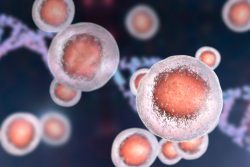 A group of researchers from Penn Engineering have discovered through novel imaging techniques that stem cells can actually control their transformation into other kinds of cells. This means that cells may have more control over their fate contrary to previous thought.
 A large scale observational study from Inserm, a French organization similar to the U.S. NIH, has found that people who consumed the most organic produce had a 25% lower risk of developing any type of cancer. While it might make sense to think all produce as equally nutritious, the growing scientific research evidence shows there is a distinct advantage to consuming organics.
 A new study has brought simple injections that can assist in regrowing damaged tissue a step closer to reality. The researchers at UBC Okanagan have created a device that makes encapsulating cells not only faster, but also more effective and cheaper.
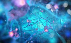 It has been the belief that mammals are born with an entire supply of neurons which have to last for a lifetime. New research has discovered a particular type of stem cell that makes neurons, which are the source of new cells in the brain’s hippocampus.
 A new study has a great new message…you can help prolong your life by increasing the power of your muscles. The study has shown for the first time that people tend to live longer lives when they have more muscle power.