Can Intermittent Fasting Help you Live Longer?
 Typically for many people a New Year is the perfect time to adopt new habits both in the gym and at the grocery store. People can be eager to try out new diets, but does scientific evidence actually support claims made for these diets?
Mark Mattson, Ph. D., a neuroscientist at John Hopkins Medicine, has studied intermittent fasting for 25 years and adopted it himself about 20 years ago. He concludes that intermittent fasting does live up to the claims that it can be a part of a healthy lifestyle.
Generally, intermittent fasting diets fall into two categories: 5:2 intermittent fasting in which people will limit themselves to one moderate sized meal two days each week, and daily time restricted eating which narrows eating times to 6 to 8 hours per day.
A variety of animal and human studies have shown that alternating between eating and fasting supports cellular health. The thought is that most likely this happens by triggering an age old adaptation to periods of food scarcity referred to as metabolic switching. This switch occurs when cells use up stores of rapidly accessible sugar based fuel and begin converting fat into energy which occurs in a slower metabolic process.
Studies have shown that this metabolic switch improves the regulation of blood sugar, suppresses inflammation, and increases the resistance to stress. Most Americans consume three meals in addition to snacks every day so they do not experience the switch and the suggested benefits.
Mattson also notes that four different studies in both people and animals found intermittent fasting also decreases blood pressure, resting heart rates, and blood lipid levels. There is also mounting evidence that this type of fasting can modify risk factors which that are associated with diabetes and obesity.
Two studies conducted at the University Hospital of South Manchester NHS Foundation Trust which involved 100 overweight women showed that the women on the 5:2 intermittent fasting diet lost the same amount of weight as women who restricted their calories. Additionally they did better with insulin sensitivity and reduced belly fat when compared to those in the reduced calorie group.
Recently, Mattson says preliminary studies have suggested that intermittent fasting could also benefit brain health. A multi center clinical trial at the University of Toronto found that 220 healthy and non obese adults who maintained a calorie restricted diet for a period of two years showed evidence of improved memory in a variety of cognitive tests. If further studies show the proof that fasting can improve learning and memory, interventions may be developed that can stave off dementia and neurodegeneration.
Mattson believes we are at a point where medical school curricula alongside advice and healthy diets and exercise may be developed. Although he recognizes that researchers do not completely understand the specific mechanisms of metabolic switching and that there are some people who or unwilling or unable to adhere to a fasting program.
With guidance and patience, most people are able to incorporate fasting regimes into their lives. It does take the body some time to adjust to intermittent fasting and to get past some of the initial hunger pangs and irritability that can accompany it. These symptoms typically pass after two weeks to a month as the brain and body become accustomed to this new habit.
To manage that hurdle, Mattson suggests that health care professionals advise patients to gradually increase the frequency and duration of the fasting periods over the course of several months, instead of going cold turkey.
To view the original scientific study click below
Effects of Intermittent Fasting on Health, Aging, and Disease.
 Researchers from the University of Illinois have conducted a new study on rats that suggests caffeine may offset some of the health risks associated with diets high in sugar and fat. What they found was that rats who consumed caffeine which was extracted from mate tea gained 16% less weight and accumulated 22% less body fat compared to rats who consumed decaffeinated mate tea.
 A new study has found that injuries to people distracted by their cell phones has increased quite steeply over the 20 year study period. People are falling, tripping, and hurting their necks and heads more often than ever. Some have even dubbed texting the “new drunk driving”!
  A new study has shown that brushing teeth frequently, three or more times per day, is linked to lower risks of heart failure and atrial fibrillation (A fib, a type of arrhythmia). Bacteria found in our mouths may be the key to many facets of our health. Some studies have found oral bacteria in blood clots of people who have had strokes, and experts have also linked gum disease to a significantly higher risk of hypertension. Mounting evidence is now strengthening the link between cardiovascular health and oral hygiene.
  By using a new method for assessing BPA levels in the human body, scientists are now suggesting that our exposure to this industrial chemical is much higher than previous estimates. Following a recent study, they believe that regulators such as the FDA could be relying on measures that have underestimated those levels by as much as 44 times.
  In new research, a team has shown a map that will identify which parts of the spinal cord trigger knees hips, ankles, and toes and the areas that put movements together. Along with an electrical spinal implant, the dream of helping people walk again could someday be a reality, even in the next decade.
 Running keeps bone marrow young! Researchers from Deakin University in Australia have found for every 5.5 miles a person runs every week, their bone marrow was one year younger.
 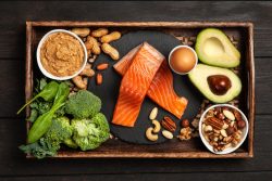 A study which involved 2,622 adults participating in the U.S. Cardiovascular Health study from 1992 to 2015, has found that higher blood levels of omega 3 fatty acids found in seafood are linked to a higher likelihood of healthy aging in older adults. With populations around the world living longer, there is a significant and growing focus on aging in a healthy manner. This means a meaningful lifespan free of major chronic diseases and with good mental and physical function.
 A pool analysis of a variety of studies and other available evidence by a group of researchers, has found that any amount of running is linked to a lower risk of early death from any cause. The researchers believe that by more people taking up running which doesn’t have to be far or fast, there would likely be remarkable improvements in population longevity and health.