
Salt Weakens the Immune System
 A current study under leadership at the University Hospital Bonn, has concluded that a diet high in salt not only is not good for blood pressure, but also for a persons immune system. Mice who were fed a diet high in salt suffered from more serious bacterial infections. Human participants who also consumed high levels of salt also showed higher immune deficiencies.
Sodium chloride which is the chemical name for salt, raises blood pressure which leads to the risk of stroke or heart attack. However, not only that – the recent study has proved for the first time that excessive intake of salt also significantly weakens an important part of the immune system.
The research team found their findings unexpected as previous studies pointed in an adverse direction. They found that infections with specific skin parasites in lab animals heal much faster if they eat a diet high in salt. The macrophages, the immune cells that attack and eat then digest parasites, are very active in salt. Many doctors concluded therefore that salt has a generally immune enhancing effect.
The results from the current study show this generalization to be inaccurate. There were two reasons for this. The body retains concentration of salt in the blood and in a variety of organs largely constant. Contrarily biological processes within the body would be impaired. Skin is the only major exception as it functions as a reservoir for salt in the body. This is the reason additional intake of salt works so well for some diseases of the skin.
However, other body parts are not exposed to the addition of salt that is eaten with food. It is filtered by the kidneys then excreted in the urine. Now the second mechanism comes into play. The kidneys have a sodium chlorine sensor that will activate the salt excretion function. As a negative side effect however, this sensor also creates so called glucocorticoids to accumulate in the body. These in turn inhibit the function of granulocytes a common type of immune cell contained in blood.
Granulocytes are scavenger cells. But they do not attack parasites, only bacteria. If they are not able to do this to a sufficient degree, infections will proceed very severely. The team was able to show this in mice that had listeria infections.
The team fed one group of mice a high salt diet and gave a normal diet to a control group of mice for comparison. In the liver and spleen of the mice that were fed a high salt diet they had counts of 100 to 1,000 increase of the number of disease causing pathogens. Listeria are bacteria that can be found in contaminated food for instance and can result in fever, vomiting and sepsis.
They also found infections of the urinary tract were found to heal much more slowly in lab mice that had been fed a high salt diet. The team traced this impaired ability to fight off bacteria infections to immune cells called neutrophils which digest bacteria. They believe the kidneys’ response to the high salt diet may indirectly affect the neutrophils.
Salt also appears to have a negative effect on the human immune system. The team examined participants who consumed six grams of salt in addition to their normal daily intake. This is about the amount contained in two fast food meals – two burgers and two servings of french fries. After one week, the team took blood from the participants and examined the granulocytes. The immune cells coped much worse with bacteria after the participants had started to consume a high salt diet.
In was also noted in the human participants, the excessive salt intake also resulted in increased glucocorticoid levels which inhibits the immune system. The best known glucocorticoid cortisone is typically used to suppress inflammation. Only through the team’s investigations in an entire organism were they able to uncover the complex control circuits that lead from salt intake to this particular immunodeficiency.
The findings are preliminary, and need larger clinical studies to confirm. However, according to the World Health Organization, a person should never consume more than 5 grams a day of salt. This amount corresponds to about one level teaspoon. Data from the Robert Koch Institute show that on average men consume 10 grams of salt per day and women 8 grams per day.
To view the original scientific study click below
A high-salt diet compromises antibacterial neutrophil responses through hormonal perturbation.
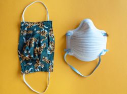 Surgical masks and N95 respirators are examples of personal protective equipment (PPE) which are used to protect the wearer from liquid contamination and airborne particles from contaminating the face. It is important to know the optimal way to prevent airborne transmission. Each type of mask and also cloth face masks serve different functions.
  A recent study by a team at the University of Leeds, UK was set up to determine if the use of paper towels or air dryers is more effective for getting rid of microbes when the hands are still drying and contanminated.
 A top exercise researcher has reported that regular exercise may reduce the risk of acute respiratory distress syndrome (ARDS) which is a major cause of death in people with the COVID-19 virus. He is now urging people to exercise regularly as a way of possibly preventing or at least reducing the severity of ARDS.
 The largest hospital system in New York is now giving high doses of Vitamin C intravenously for patients with COVID 19. The treatment is based on Chinese reports of this approach helping patients with the virus.
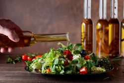 The team at the University of Minnesota Medical School researched olive oil and may have found what it is that activates the pathway to increased lifespan and health!
 How we balance weight gain and weight loss is predominantly due to what we eat, how much we eat and by how much we exercise. However, another important factor is often looked over. It isn’t just about the amount of calories we consume, but when they are consumed that will determine how well we will burn those calories.
 A new large study has linked consuming processed meats and red meats to a higher risk of death and heart disease. The study from Northwestern Medicine and Cornell University refutes a previous controversial study concluded that it was not needed for people to alter their diet in regards to processed and red meats.
 New research has indicated that eating a Mediterranean diet for a year can help boost gut bacteria which are linked to healthy aging. The five country study also indicates this diet can help reduce bacteria that is associated with dangerous inflammation people that are older.
 According to a new study led by Sylvia Santosa at Concordia University’s Faculty of Arts and Sciences, the mechanisms by which aging and obesity develop are very similar. The team believes that obesity should now be considered premature aging because it predisposes people to acquiring potentially life altering diseases which are normally seen in older people.