Loss of Taste and Smell Linked to Higher Chance of Covid-19

Both the loss of taste and smell have been anecdotally linked to a higher chance of developing the COVID-19 virus. The recent study has shown people are more than 10 times more likely to develop the virus infection than any other causes of infection.

Researchers at UC San Diego Health have reported that the first empirical findings strongly associate sensory loss with COVID-19. The team surveyed 1,480 patients who had flu-like symptoms and concerns regarding the potential of infection. Contained in that total, 102 patients tested positive for the virus and 1,378 tested negative. The study included responses from 203 COVID-19 negative patients and 59 positive patients.

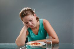
68% of the positive participants reported loss of smell and 71% reported loss of taste as compared with 16% and 17% of negative patients respectively. None of the participants in the study required hospitalization or invasive breathing support. However, if social distancing was not in place, these individuals could possibly spread the infection to others within their community despite not experiencing severe symptoms.

The study has demonstrated the unique presentation and high prevalence of certain sensory impairments in patients who were positive with the virus. Of those patients who reported loss of taste and smell the loss typically profound and not mild. However, encouragingly the rate of recovery of taste and smell was high and typically occurred within two to four weeks after infection.

The study not only showed that the high occurrence of taste and smell loss is specific to the COVID-19 virus, but also fortunately has shown that the majority of people experienced sensory recovery fairly rapidly. Within the COVID-19 patients with loss of smell, more than 70% reported improvement in smell at the time of the survey. Most of those who hadn’t reported improvement had only been diagnosed recently.

The return of taste and smell typically matched the timing of recovery from the disease. Interestingly, the researchers found that people who reported experiencing a sore throat more often tested negative for the virus.

To help decrease the risk of virus transmission, UC San Diego Health now includes loss of taste and smell as a screening requirement for staff and visitors. The screening is also a marker for testing patients who might be positive for the virus.

Other symptoms of COVID-19 include fatigue, fever, difficulty breathing, and cough. Respondents in the study were most often people with milder forms of COVID-19 infection who did not require hospitalization or intubation. The team’s findings stress the importance of identifying subtle and early symptoms of the virus in people who might be at risk of transmitting the disease as they recuperate within their community.

The CDC has officially listed loss of taste and smell as symptoms of the virus which is a validation of the ruminating concerns about these symptoms around the world. It is the hope that the team’s findings inspire other institutions to follow suit and not only list taste and smell loss as a symptom of the virus, but also use it as a screening measure for the virus around the world.

To view the original scientific study click below

Association of chemosensory dysfunction and Covid-19 in patients presenting with influenza-like symptoms.

Does Sleep Loss Cause Obesity?

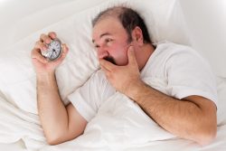
New research has found that quite the opposite is true. Previously a growing body of research has suggested that a poor quality of sleep is linked to an increased risk of obesity through deregulating appetite which results in higher caloric consumption. However, the latest study actually shows a flip side to that suggesting obesity leads to poor sleep. It isn’t the loss of sleep that leads to obesity, but instead it is excess weight that can lead to poor sleep.

It is thought that sleep is a function of the body trying to conserve energy where energetic levels are going down. The recent studies findings suggest that if a person were to fast for a day, they would get sleepy because their energetic stores would be depleted.

In humans, acute disruption of sleep can result in an increase in appetite and also insulin resistance. People who chronically get less than 6 hours of sleep per night are more likely to be diabetic and obese. However, starvation in rats, worms, fruit flies and humans has been shown to affect sleep which indicates that it is regulated at least in part by the availability of nutrients. However, the ways in which eating and sleeping work in tandem has been unclear.

The study team wanted to know what is sleep actually doing? Short periods of sleep and other chronic conditions such as diabetes, are linked but it is just an association. It isn’t clear if short sleep is causing the propensity for obesity or instead that the obesity itself causes the propensity of short sleep.

To research the association between sleep and metabolism, the team genetically altered C elegan worms to turn off a neuron that controls sleep. These worms could still breathe, eat and reproduce, but lost their ability to sleep. With the neuron disabled, the team saw a severe drop in adenosine triphosphate (ATP) levels which is the body’s energy currency.

In earlier research, a gene in C elegans called KIN-29 was studied. This particular gene is homologous to the Salt Inducible Kinase (SIK-3) gene found in humans. This gene had already known to promote sleep pressure. When the researchers knocked out the KIN-29 gene to promote sleepless worms, the mutant C elegans amassed more fat which resembles the human condition of obesity, although their ATP levels were decreased.

The team hypothesized that releasing fat stores is a control by which sleep is encouraged and the reasons KIN-29 mutants could not sleep was because they were not able to release the fat. By testing this, the team again manipulated the KIN-29 mutant worms by releasing an enzyme that discharged their fat. Because of this manipulation, once again the worms were able to sleep.

The team said this could explain a reason why people who are obese may experience sleep problems. There may be a signaling problem between the brain cells that control sleep and fat stores.

There is still much to unravel about sleep, however the new research provides a step closer to understanding sleep’s core functions and how to treat common sleep disorders. There appears to be a common overarching sentiment within the sleep field that sleep is all about the brain or the nerve cells. The new work suggests that may not be true. There are complex interactions between the brain and the rest of the human body that connects to regulation of sleep.

To view the original scientific study click below

A salt-induced kinase is required for the metabolic regulation of sleep.

Vitamin D Supplementation may Reduce Risk of Both the Flu and Covid-19

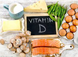
With the world in the middle of the COVID-19 pandemic, public health policies that could decrease the danger of infection and even death along with less quarantines are greatly needed. Vitamin D supplementation shows promise in reducing those risks in both the COVID-19 virus and influenza.

Through a variety of mechanisms, Vitamin D could decrease the chance of infections. These mechanisms such as inducing defensins and cathelicidins which are both cysteine rich cationic proteins that play a critical role in mammalian innate immune defense against invasive bacterial infections. These proteins may decrease viral replication rates and reduce concentration levels of pro-inflammatory cytokines that cause inflammation that damages the lung linings and may lead to pneumonia and increasing concentration levels of anti-inflammatory cytokines also.

Vitamin D is a modulator of adaptive immunity. It suppresses responses mediated by the T help cell type by primarily repressing production of inflammatory cytokines. Serum concentrations tend to decrease with age which may be critical to COVID-19 because case fatality rates increase with age.

Several clinical trials and observational studies have reported that taking Vitamin D decreased the risk of influenza, and others could not. Supporting evidence of the role Vitamin D may play in reducing the risk of COVID-19 includes that the outbreak resulted in the winter which is a time when
25-hydroxyvitamin D (25(OH)D) concentrations are at their lowest. The amount of cases in the Southern Hemisphere at summers end are low. Additionally it has been found that Vitamin D deficiency may promote acute respiratory distress syndrome and that fatality rates can increase as a person ages and with extreme disease comorbidity, which both are associated with decreased (25(OH)D) concentration.

To minimize the promotion of infection, it has been recommended that if you are at risk of influenza and/or COVID-19 would maybe take 10,000 IU/d of Vitamin D3 for a couple of weeks to rapidly increase 25(OH)D) levels followed by 5,000 IU/d. The goal is to increase 25(OH)D) levels of concentrations above 40-60 ng/mL. For the treating people that have already become infected with COVID-19, higher levels of Vitamin D3 doses could be useful.

Magnesium supplementation is recommended when taking Vitamin D supplements as it helps activate Vitamin D which in turn promotes regulation of calcium and phosphate homeostasis to affect maintenance and growth of bones. All enzymes which will metabolize Vitamin D appear to need Magnesium, acting as a cofactor in the enzymatic reactions of the kidneys and liver. The dose of Magnesium should be in the range of 250-500 mg/d along with twice the dose of calcium.

Although some contradictory data exists, the evidence available shows that supplementation with several micronutrients with immune supporting roles can vary immune function and help decrease the risk of infection. Micronutrients that haveh the strongest indications for immune support are Vitamins D and C along with Zinc.

To view the original scientific study click below

Evidence that Vitamin D Supplementation Could Reduce Risk of Influenza and COVID-19 Infections and Deaths.

Stem Cell Technology Rejuvenates Old Human Cells

According to a new study by researchers at Stanford University of Medicine, old human cells will return to a more vigorous and youthful state following being induced to briefly express a protein panel involved in embryonic development. They team also discovered that elderly mice regained their youthful strength after their existing muscle stem cells were subjected to the protein treatment rejuvenation and then transplanted back into their bodies.

The proteins which are known as Yamanaka factors are typically used to transform adults cells into induced pluripotent stem cells or iPS cells. These iPS cells can become nearly any type of cell within the body regardless of the cell from which they originated. They have become important in regenerative medicine and drug discoveries.

The current study found that inducing old human cells in a lab dish for a brief period to express these proteins rewinds many of the molecular hallmarks of aging. They render the treated cells nearly indistinguishable from their younger counterparts.

When adult cells make iPS cells they become pluripotent and youthful. The team wondered if it might be possible to just rewind the aging clock without inducing pluripotency. They found that by tightly controlling the duration of the exposure to these protein factors, they were able to promote rejuvenation in multiple types of human cells.

The researchers make iPS cells from adult cells such as those that compose skin through repeatedly exposing them over a two week period to a panel of proteins which are vital to early embryonic development. They do so by introducing daily short lived RNA messages into the adult cells. The RNA messages then encode the instructions for making the Yamanaka proteins. These proteins over time rewind the fate of the cell by pushing them backward along the timeline of development until they resemble young, embryonic like pluripotent cells from which they originated.

During the process the cells shed any memories of their previous identities and they revert to a younger state. They accomplish this by wiping their DNA clean of the molecular tags that not only differentiate for example a skin cell from a heart muscle cell, but of other tags that accumulate as cell age.

The team wondered if old human cells would respond similarly and whether the response would be limited to a few cell types or generalizable for many tissues. They created a way to use genetic material called messenger RNA to temporarily express six reprogramming factors which were the four Yamanaka factors plus two additional proteins found in blood vessel cells and human skin. Messenger RNA will rapidly degrade in cells which allowed the researchers to tightly control the duration of the signal.

The team then compared the gene expression pattern of the treated cells and control cells which were both obtained from older adults, with those of untreated cells from younger people. They discovered that cells from the older adults exhibited signs of aging reversal following four days of exposure to the reprogramming factors. However the untreated older cells expressed higher levels of genes associated with known aging pathways. Treated older cells more closely resembled younger cells in their patterns of gene expression.

When they studied the patterns of aging associated chemical tags which are known as methyl groups which serve as indicators of a cell’s chronological age, they discovered that the treated cells appeared to be approximately 1-1/2 to 3-1/2 younger on average than the untreated cells from the older people. They showed peaks of 3-1/2 years (in skin cells) and 7-1/2 years in cells that line blood vessels.

The researchers then compared several hallmarks of aging including how cells metabolize compounds to create energy and dispose of cellular trash and how cells sense nutrients, among cells from young people, treated cells from older adults and untreated cells fro older adults.

They saw a dramatic rejuvenation across all the hallmarks but one in all the types of cells that were tested. The last and most experiment was done stem cells found in the muscles. Although these cell types are naturally endowed with the ability to self renew, this ability wanes with aging. The team wondered if they could also rejuvenate these stem cells and have a long term effect?

When they transplanted elderly mouse muscle stem cells that had been treated back into elderly mice, the mice regained the muscle strength similar to younger mice.

The team then isolated cells from the cartilage of people without and with osteoarthritis. They discovered that the temporary exposure of the osteoarthritic cells to the reprogramming factors reduced the secretion of inflammatory molecules and also improved the ability of the cells to divide and function.

The team is now optimizing the panel of reprogramming proteins needed to rejuvenate human cells. They are also looking at the possibility of treating tissues or cells without removing them from the body.

To view the original scientific study click below

Transient non-integrative expression of nuclear reprogramming factors promotes multifaceted amelioration of aging in human cells.

Best Materials for Homemade Face Masks

During the beginning of the COVID-19 Pandemic CDC recommends people wear face masks when in public. Due to the shortage of N95 and surgical masks, they have recommended people make their own coverings. Some materials can filter out particles much better than others, significantly affecting how well a homemade face mask may protect a person from the virus.

The COVID-19 virus is believed to be spread mainly from respiratory droplets from a person that’s been infected speaks, breathes, coughs or sneezes. The droplets are all different sizes, however the tiniest ones, which are called aerosols can slip through any openings that are between a variety of cloth fibers. This has lead to some people questioning as to whether cloth masks can really help fight disease.

Supratik Guha at the Univ. of Chicago and colleagues set out to study if common fabrics by themselves or in a combination have the ability to filter out aerosols in relation in size to respiratory droplets.

The team used an aerosol mixing device to create particles that range from 10nm to 6um in diameter. A fan was used to blow the aerosol across a variety of cloth samples at an airflow rate similar to a person’s respiration at rest. The team then measured the size and number of particles in air before and following passing through the fabrics.

Tightly woven cloth in one layer along with 2 layers of polyester-spandex chiffon which is a sheer fabric, filtered out most of the aerosol particles ((80-99%) depending on the size of the particle with results close to that of an N95 mask.

They then substituted using natural silk or flannel or just using cotton quilt with cotton-polyester batting which had similar results. The team has pointed out that tightly woven fabrics like cotton man act as a mechanical border to particles. Fabrics that hold a static charge such as natural silk and chiffon act as an electrostatic barrier.

Researchers are reporting that a combination of cotton with either natural silk or chiffon can effectively filter out aerosol particles if the fit is good. You can also use doubled up 600 thread count pillowcases or flannel pajamas or cotton T-shirts which will make a mask that will provide up to 60% filtration. Other materials that can be used to filter our dangerous particles include vacuum cleaner bags, HEPA filters, and quilter’s cotton. You do want to be careful of materials like household air filters that many contain fiberglass which can damage the lungs even if they filter out particles.

To view the original scientific study click below

Aerosol Filtration Efficiency of Common Fabrics Used in Respiratory Cloth Masks.

Salt Weakens the Immune System

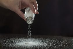
A current study under leadership at the University Hospital Bonn, has concluded that a diet high in salt not only is not good for blood pressure, but also for a persons immune system. Mice who were fed a diet high in salt suffered from more serious bacterial infections. Human participants who also consumed high levels of salt also showed higher immune deficiencies.

Sodium chloride which is the chemical name for salt, raises blood pressure which leads to the risk of stroke or heart attack. However, not only that – the recent study has proved for the first time that excessive intake of salt also significantly weakens an important part of the immune system.

The research team found their findings unexpected as previous studies pointed in an adverse direction. They found that infections with specific skin parasites in lab animals heal much faster if they eat a diet high in salt. The macrophages, the immune cells that attack and eat then digest parasites, are very active in salt. Many doctors concluded therefore that salt has a generally immune enhancing effect.

The results from the current study show this generalization to be inaccurate. There were two reasons for this. The body retains concentration of salt in the blood and in a variety of organs largely constant. Contrarily biological processes within the body would be impaired. Skin is the only major exception as it functions as a reservoir for salt in the body. This is the reason additional intake of salt works so well for some diseases of the skin.

However, other body parts are not exposed to the addition of salt that is eaten with food. It is filtered by the kidneys then excreted in the urine. Now the second mechanism comes into play. The kidneys have a sodium chlorine sensor that will activate the salt excretion function. As a negative side effect however, this sensor also creates so called glucocorticoids to accumulate in the body. These in turn inhibit the function of granulocytes a common type of immune cell contained in blood.

Granulocytes are scavenger cells. But they do not attack parasites, only bacteria. If they are not able to do this to a sufficient degree, infections will proceed very severely. The team was able to show this in mice that had listeria infections.

The team fed one group of mice a high salt diet and gave a normal diet to a control group of mice for comparison. In the liver and spleen of the mice that were fed a high salt diet they had counts of 100 to 1,000 increase of the number of disease causing pathogens. Listeria are bacteria that can be found in contaminated food for instance and can result in fever, vomiting and sepsis.

They also found infections of the urinary tract were found to heal much more slowly in lab mice that had been fed a high salt diet. The team traced this impaired ability to fight off bacteria infections to immune cells called neutrophils which digest bacteria. They believe the kidneys’ response to the high salt diet may indirectly affect the neutrophils.

Salt also appears to have a negative effect on the human immune system. The team examined participants who consumed six grams of salt in addition to their normal daily intake. This is about the amount contained in two fast food meals – two burgers and two servings of french fries. After one week, the team took blood from the participants and examined the granulocytes. The immune cells coped much worse with bacteria after the participants had started to consume a high salt diet.

In was also noted in the human participants, the excessive salt intake also resulted in increased glucocorticoid levels which inhibits the immune system. The best known glucocorticoid cortisone is typically used to suppress inflammation. Only through the team’s investigations in an entire organism were they able to uncover the complex control circuits that lead from salt intake to this particular immunodeficiency.

The findings are preliminary, and need larger clinical studies to confirm. However, according to the World Health Organization, a person should never consume more than 5 grams a day of salt. This amount corresponds to about one level teaspoon. Data from the Robert Koch Institute show that on average men consume 10 grams of salt per day and women 8 grams per day.

To view the original scientific study click below

A high-salt diet compromises antibacterial neutrophil responses through hormonal perturbation.

Unmasking the Differences between Face Masks

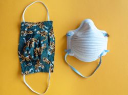
Surgical masks and N95 respirators are examples of personal protective equipment (PPE) which are used to protect the wearer from liquid contamination and airborne particles from contaminating the face. It is important to know the optimal way to prevent airborne transmission. Each type of mask and also cloth face masks serve different functions.

N95 respirators are tested and approved by the NIOSH (National Institute for Occupational Safety and Health). Their intended use and purpose is to protect the wearer from exposure to particles including small particle aerosols and large droplets. They are designed to have a close fit to the face and very effective filtration of particles that are airborne.

The N95 classification means that when exposed to careful testing, the respirator prohibits a minimum of 95% of quite small (0.3 micron) particles that were tested. If fitted properly, the filtration properties of these type respirators exceed those of face masks. However, they do not completely eliminate the risk of illness. Leakage can occur around the edge of the mask when user inhales. N95 respirators are disposable and ideally should be discarded after each patient encounter and/or when the unit no longer forms an effective seal to the face, becomes wet or visibly dirty, breathing becomes difficult, or it becomes contaminated with blood, respiratory or nasal secretions or other bodily fluids from patients.

N95 masks are typically used in healthcare settings. The CDC doesn’t recommend to the public to wear N95 respirators to safeguard themselves against respiratory diseases such as COVID-19. These masks are critical supplies that are primarily reserved for health care workers and first responders as recommended by the CDC.

Surgical masks are disposable, loose fitting accessory that devise a physical barrier between the nose and mouth of the wearer and potential contaminants in the immediate environment. They are typically used for the protection of the wearer from splashed sprays and large particle droplets. They also prevent the spread of possibly infectious respiratory secretions from the wearers to others. They are tested and approved by the FDA.

Surgical masks are loose fitting and can vary in design. However, masks are often flat, rectangular and have folds and/or pleats. The top of the mask can contain a metal piece which can be bent to a person’s nose. Long straight ties or elastic bands help a surgical mask stay in place while being worn – either looped behind the ears or tied behind the head.

While a surgical mask is meant to block large particle splashes, droplets, splatter or sprays that might contain germs (bacteria and viruses), they cannot block or filter particles that are very small in the air that could be carried by sneezes, coughs or certain medical procedures. They do not achieve complete protection from other contaminants and germs because they have a loose fit between the layer of the face mask and face. They are intended for one use only and should be discarded after each patient encounter.

Cloth face coverings many of which can be homemade, only offer a small degree of protection, but the may also help the slow spread of the COVID-19 virus. These type masks are what the CDC is recommending for use by the general public when in public settings.

Face cloths should not be placed on children under the age of 2, people who have breathing difficulties, people who are unconscious, or people who are unable to remove the mask on their own. They should be washed after each use. When removing a person should be careful not to touch their nose, eyes or mouth and hands should be washed immediately after removal.

They can be made from common materials. It should be kept in mind by wearers of these type masks that they can provide a false sense of security. While they do offer some degree of protection, they offer a lot less protection than respirators or surgical masks. One study indicated that homemade face masks may be half as effective as surgical masks and up to 50 times less effective than N95 respirators. However, they are better than not wearing any mask and create some protection especially where a person cannot social distance.

Removing Virus After Hand Washing – Paper Towels vs. Air Dryers

A recent study by a team at the University of Leeds, UK was set up to determine if the use of paper towels or air dryers is more effective for getting rid of microbes when the hands are still drying and contanminated.

Hand drying is especially critical in helping decrease the spread of critical microbes which includes COVID-19. Failure to remove these microbes increases their transmission to environmental surfaces which increases the opportunity for them to be transmitted and spread. The study set out to investigate if there could be differences in the extent of transmission of the virus according to each hand drying method outside of the bathroom to a hospital environment.

Four participants simulated contamination of either their hands or gloved hands with a bacteriophage, a virus that can infect bacteria and is not harmful to humans. They did not wash their hands after contamination as the method used to simulate poorly and inadequately hand washing. They dried their hands using either a jet air dryer or paper towels. Each participant did wear an apron to ensure measurement of contamination of body/clothing while their hands dried. They dried their hands in a hospital bathroom and after the participants exited, samples were retrieved from both ward and public areas.

Environmental surface areas were sampled after the participant’s contact with hands or apron. These site samples included doors, stair handrails, chairs in ward and public areas, lift buttons, phones, buttons on intercoms to wards, stethoscope tubing and chest and head piece, the aprons, and armchairs that might have been indirectly exposed to the aprons. For the latter sample site, the participants were told to lay their arms across their chest while using the apron and before resting their arms on the chair.

The study team discovered that both paper towel and air dryer methods statistically significantly decreased virus contamination of hands. For 10 out of 11 areas tested, quite a high amount of environmental contamination was found after air dryers versus paper towel use. All surfaces sampled following the use of air dryers showed phage contamination compared to 6 surfaces after paper towel use. Average surface contamination following hand contact was more than 10 times higher after air dryer use versus paper towel use.

Viral dispersal to apron/clothing was 5 times higher with air dryers versus the paper towels. Phage transfer from aprons to armchairs via the crossed arms was detected only following air dryer use. This suggests that the transfer of microbes to environmental surfaces can occur directly from hands that remain contaminated after air drying, but also indirectly from a person’s body that has been contaminated during air drying.

The research team concluded that there are very distinct differences in regards to air drying versus paper towel drying in the residual microbial contamination of the participant’s body and hands. These differences in contamination translate into quite a bit greater microbe levels of contamination after air drying versus the use of paper towels from body and hands beyond the bathroom. Since public bathrooms are used by staff, visitors and patients, the hand drying method used can promote the increase (using air dryers) or decrease (using paper towels) pathogen transmission in hospitals.

The team also notes that their findings have significant importance since there has been a general migration from use of paper towels to more air drying in many settings and areas in the world. The World Health Organization’s hand washing guidelines recommend the use of paper towels to dry hands. And they also recommend using a paper towel to turn off the tap. The preferred method should be paper towels to dry hands following proper washing to help reduce the risk of virus contamination and spread.

To view the original scientific study click below

Dispersal of microbes to hospital surfaces following two hand drying methods: paper towels or a jet air dryer

Protect Against Deadly Covid-19 Complications with Exercise

A top exercise researcher has reported that regular exercise may reduce the risk of acute respiratory distress syndrome (ARDS) which is a major cause of death in people with the COVID-19 virus. He is now urging people to exercise regularly as a way of possibly preventing or at least reducing the severity of ARDS.

Zhen Yan, PhD at the University of Virginia School of Medicine has shown that findings from medical research strongly supports the possibility of exercise as a form of treatment for those suffering from ARDS. Between 3% and 17% of all patients with the COVID-19 virus suffer from ARDS. The Centers for Disease Control and Prevention has estimated that 20% to 42% of patients who become hospitalized with COVID-19 will develop ARDS. For patients admitted to an ICU the range is estimated at 67% to 85%.

Research that was conducted prior to the COVID-19 pandemic has suggested that about 45% of patients who develop severe ARDS will die. Right now what we hear is that social distancing or ventilators is all we have to avoid exposure or relying on a ventilator to survive the virus. However, 80% of confirmed COVID 19 patients have only mild symptoms with no need for respiratory support and the question is why.

Research findings in regards to an endogenous antioxidant enzyme may provide important clues. Extracellular Superoxide Dismutase (EcSOD) is a potent antioxidant that hunts down harmful free radicals which helps protect our tissues and prevent disease. Our muscles naturally produce this antioxidant secreting it into circulation to allow binding to other vital organs. Its production is enhanced by cardiovascular exercise.

A decrease in EcSOD is seen in a variety of diseases including ischemic heart disease, kidney failure and acute lung disease. Research conducted on mice suggests that blocking this antioxidant production worsens heart problems, while increasing its production has beneficial effects. A decrease in EcSOD is also associated with chronic conditions like osteoarthritis.

Research has suggested that even a single session of exercise increases the production of EcSOD. With the current COVID-19 pandemic Dr. Yan is urging people to find ways to exercise while still maintaining social distancing. Regular exercise has far more health benefits that we currently know. The protection exercise may provide against ARDS which is a serious condition that can develop in COVID 19 patients, is just one of the many examples.

Dr. Yan’s review has strongly suggested EcSOD has a potential and promising treatment for ARDS and a variety of other health conditions. Gene therapy is another example which may one day be used to increase the production of EcSOD so that its protection presence in lungs is enhanced in patients who are battling the COVID-19 virus.

Research has also shown that rats with chronic kidney disease had less damage to their kidneys when treated with human EcSOD. This antioxidant is being proposed as a potential therapy for diabetic retinopathy which is a complication of diabetes that can lead to blindness.

EcSOD may also prove beneficial for multi-organ dysfunction syndrome where multiple organs begin to fail. Treating the condition with general antioxidants has not been successful, however Dr. Yan suggests that understanding EcSOD’s workings may lead to doctors using it in a more targeted and more effective fashion.

It is often said that exercise is medicine. EcSOD has set a perfect example of the biological process of exercise to potentially advance medicine.

To view the original scientific study click below

Extracellular superoxide dismutase, a molecular transducer of health benefits of exercise.

High Dose Intravenous Vitamin C for Covid-19

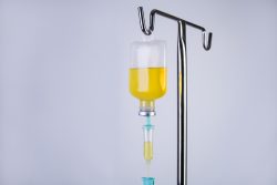
The largest hospital system in New York is now giving high doses of Vitamin C intravenously for patients with COVID 19. The treatment is based on Chinese reports of this approach helping patients with the virus.

Critical care specialist and pulmonologist Dr. Andrew G. Weber has reported that his COVID 19 patients in intensive care are receiving 1,500 mgs of Vitamin C intravenously and are being re-administered the dose 3 to 4 times per day. These doses are 16 times more than the national recommended allowances of 75 mg for adult women and 90 mg for adult men. The approach is based on experimental treatments and small studies with COVID 19 patients in Shanghai, China.

The information from China describes patients with new coronaviral pneumonia who recovered and were discharged from the hospital after high dose Vitamin C. The information also states that early application of large doses of Vitamin C can have a strong antioxidant effect and improve endothelial function. High dose Vitamin C has been shown to not only improve antiviral levels but also can prevent and treat acute respiratory distress and acute lung injury.

Vitamin C is being widely used to treat patients with COVID 19 throughout the system. The treatment protocols do vary from patient to patient. Currently more COVID 19 patients are being treated in the hospital network in New York than anywhere else in the United States.

The Vitamin C treatments are being given along with other medicines including hydroxychloroquine, azithromycin and other biologicals and blood thinners. These “cocktails” are being administered to severely ill patients on a compassionate care basis. The patients who received the additional Vitamin C showed significant improvement over those who did not.

Vitamin C levels drop dramatically in people with COVID 19 when they experience sepsis. Sepsis is an inflammatory response that occurs when the body overreacts to an infection. It therefore makes sense to try and maintain levels of Vitamin C.

A hospital in Wuhan, China is currently conducting clinical trials in regards to the effectiveness of high dose intravenous Vitamin C on COVID 19 patients.