 As we search for the fountain of youth, fecal transplants might seem to be an unlikely method in the process of reversing aging. However, researchers have shown evidence from their study on mice, that fecal microbiota transplanting from young mice into older mice may reverse signs of aging in the brain, eyes, and gut.
In the study, microbes from older mice created inflammation in young recipient’s brains and reduced an important protein which is needed for correct vision. The discoveries have shown that gut microbes have a part in the regulation of some negative effects of aging and opens up the opportunity of therapies that are gut microbe based to combat the decline in older life.
The ground breaking work provided exciting realizations for the involvement of microbes in the gut in aging and the decline of brain and vision function. It also provides a possible solution from replacement therapy with gut microbes.
It is known that the microbe population that people carry in the gut, which is collectively known as gut microbiota, is associated with health. Many diseases are linked to changes in the behavior and types of viruses, fungi, bacteria and other microbes in a person’s gut.
Some changes in the composition of the microbiota occur as people age, negatively affecting immunity and metabolism. This can be directly linked to age related disorders such as cardiovascular, inflammatory bowel disease, metabolic, neurodegenerative and autoimmune disorders.
In understanding the results of the changes as we age in the microbiota, the team transferred gut microbes from older mice into young healthy mice and vice versa. Then they observed what affects it had on inflammation of aging in the brain, vision and gut which all suffer from the decline of function as a person gets older.
This study discovered that the microbiota from the older donor mice led to a loss of stability of the gut lining. This allowed bacterial products to incorporate into circulation, which caused inflammation in the eyes, brain and the immune system.
Inflammaging which is chronic age related inflammation, has been linked with activation of very specific immune cells that are in the brain. These particular cells became greatly activated in the younger mice who had received microbiome transplants that were aged.
The researchers additionally noted specific proteins in the eye linked with degeneration of the retina that were at higher levels in the younger mice who had received microbiota from the older donors.
In aged mice, these negative developments in the eye, brain and gut could be restored through transplanting gut microbiota from the younger mice.
In continuing studies, the researchers are in the process to try and understand the long term effects that were positive can last. They also hope to identify the components that are beneficial of the young mice microbiota and what the impact is on organs that are far off from the gut.
The microbiota of the younger mice, and the older mice who had been given younger transplants of microbiota, were fortified in bacteria that was beneficial and had previously been linked with good health in both humans and mice.
The team also analyzed the products that these bacteria are producing by breaking down certain elements of what we eat. This has shown major changes in certain fats (lipids) and metabolism of vitamins which could be associated to the shifts that are seen in cells that are in the brain and eyes.
The same pathways live in people and the gut microbiota of humans also will change greatly as we get older, but the team caution that extrapolating the results of their research to humans until studies that are similar in older people can be made.
A new facility for MRT (Microbiota Replacement Therapy) which is also known as FMT (Fecal Microbiota Transplantation) is being built that will enable these trials in addition to new trials for conditions that are microbiota related.
The team was excited to discover that through changing the gut microbiota of older people, they could rescue indicators of age related decline which is commonly seen in conditions that are degenerative in the eye and brain.
The results provide additional evidence of the major links between gut microbes and the healthy aging of organs and tissues of the body. They hope their findings ultimately contribute to understanding how manipulation of diet and gut bacteria can maximize better health as we age.
Fecal transplants are relatively safe and rarely spread disease as long as they are lab tested for diseases and are from very healthy donors. At one time doctors could order them for their patients, however the FDA in the US now requires extreme illness before they are allowed. This is sad since side effects and complications have been rare.
To view the original scientific study click below:
Fecal microbiota transfer between young and aged mice reverses hallmarks of the aging gut, eye, and brain

 Loss of hearing due to noise, certain cancer drugs and aging cannot be reversed due to researchers not being able to reprogram cells to evolve into the inner and outer ear sensory cells. This is fundamental for hearing after they have died. However, researchers have now found a particular master gene to program ear hair cells into an inner or outer cell. This overcomes a significant hurdle that has prevented these cells from developing and hearing to be restored.
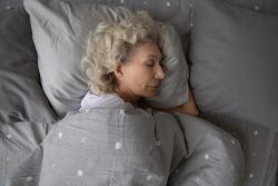 A new study has shown that seven hours of sleep a night is optimal for those in their mid to older ages. And, too much or too little sleep is linked with poorer mental health and cognitive performance.
  Close to half of older adults might have sleep apnea, which is a condition where sleep and breathing are briefly interrupted many times throughout the night. A recent study has shown that this chronic tiredness may have serious implications for safety on the road.
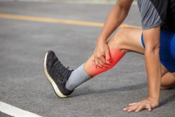 Many people know that an injury to tendons can be difficult, lengthy and many times an incomplete process of healing. As an example, repetitive or sudden motion which can be experienced by factory workers and athletes can increase the risk of ruptures or tears in the tendons. 30% of people will experience an injury to the tendon with the highest risk in women. Additionally, people who endure this type of injury are more likely to experience a future injury at the same site or never fully recover from the injury.
 Every day we all make decisions about what we consume, however our choices might not be totally our own. Research on mice has shown that the microbes in their guts have an influence on what they choose to eat, creating substances that will prompt urges for a variety of foods.
 If you make mistakes or are forgetful when hurried, a new study which is the largest in this field to date, has found that meditation can help a person be less error prone.
 Vitamin D is naturally produced when you are exposed to the sun. It is a natural source of a hormone essential to us and especially to our bones. However, according to new research, when you are low on this vitamin, not only do the bones take a hit but also cardio health.
 Researchers have cultivated successfully human stem cells that have the ability to renew themselves and repair damage to muscle tissue in mice. This potentially advances attempts to treat disorders that are muscle wasting and muscle injuries in humans.
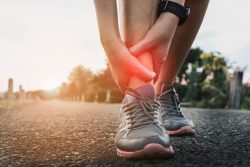 Researchers have developed a promising new approach to combat age related muscle atrophy that is associated with immobility following illness or injury. The technique which was shown in mice, arrests the processes through which muscle will begin to deteriorate at the start of exercise following a period of inactivity.