
Combination Therapy Boosts Spinal Cord Injuries
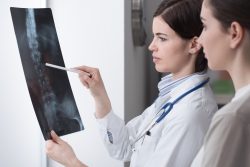 Intensive physical therapy and stem cell grafts together can boost the functionality of spinal cord injuries more than either treatment alone. Researchers found this in animal models, where tissue growth, repair, and functionality were increased following the combination of therapies. The stem cells promote growth and healing of the surrounding tissues, while the physical therapy helps to improve movement and function.
Implants of stem cells or neural grafts can promote regeneration in spinal cord injuries when used with intensive physical rehabilitation. This combination may help improve function by promoting new or greater roles for spared or undamaged cells or neural circuits.
Stem cells have the ability to promote physical and functional recovery in individuals who have experienced a spinal cord injury. A recent study showed that rats who had a cervical lesion and then received a neural stem cell graft had improved functionality and movement when compared to rats who only received the lesion or physical therapy.
It was found that rehabilitation therapy for the animals begun one month following the injury helps approximate when human patients are admitted to spinal cord injury rehabilitation centers. In addition, this therapy rewards animals for grasping skills, which ultimately promotes their rehabilitation. Physical therapy can help promote regeneration of injured spinal cord nerve cell and, additionally, stem cell grafts may also be effective in aiding recovery. When both treatments are administered one month following the injury, there is significant improvement in grasping ability.
The new findings show that rehabilitation plays a critically important role in helping people with spinal cord injuries recover function. When combined with pro-regenerative therapies, such as stem cell transplants and physical rehabilitation it can significantly improve outcomes. This is surprising, as the benefits of physical rehabilitation were found to be much greater than what has been observed in humans. This suggests that early and intense rehabilitation may be key to maximizing functional recovery after a stem cell transplant.
Spinal cord injuries are a medical challenge that is still unresolved for many people. Each year, nearly 18,000 people in the U.S. suffer from an injury to the spinal cord. This often leads to some degree of physical impairment or paralysis that is permanent.
There is a need to improve therapies following spinal cord injuries. Researchers hope that this new combination therapy can help improve the function of the spine after an injury. Clinical human trials are planned for the near future to test this theory.
To view the original scientific study click below:
Rehabilitation combined with neural progenitor cell grafts enables functional recovery in chronic spinal cord injury
 If you’re looking to improve your muscular strength without the need for exercise, then this is good news! Green leafy vegetables are a great source of nitrates, and just one cup per day can provide a significant boost in muscle function.
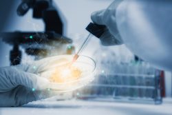 A team of researchers have discovered some key contributors that encourage human stem cell reprogramming to the naive state. This can be utilized to model early stages of development and will help scientists generate naive pluripotent stem cells quickly and efficiently. The discovery will help provide new understanding into the systems that reconfigure and destabilize cell identity involved in transitioning states of cells. The team learned more about reprogramming of naive stem cells after a genome wide function screen.
 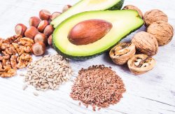 Heart disease is a leading cause of death in the United States, so it’s important to know which foods can help reduce the risk. A new research review has found that the major plant-based version of the nutrient omega-3 fatty acid, alpha-linolenic acid (ALA), can benefit heart health and reduce the risk of heart disease.
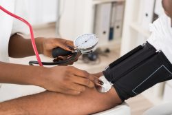 Recent research has found that mice with high blood pressure lose more bone than those without the condition. As humans age, their bones become weaker and more brittle as a result of chronic inflammation which can lead to osteoporosis-related fractures when not treated properly. The team suggests treatments for this type of hypertension throughout early adulthood might help prevent further damage during later years. However, they also say it’s important we find out how prevalent these traits are among younger individuals so treatment options exist if needed.
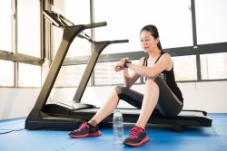 Getting more than the minimum recommended amount of exercise could help you live a healthier and longer life according to a new study. The current guidelines recommend 75 to 300 minutes of exercise on a weekly basis to benefit health. By doing more, you are linked to even a lower risk of issues with heart health and other risk factors. Up to 5 hours of exercise that is vigorous and 10 hours of moderate exercise such as walking may help decrease the risk of early death.
 A new study by researchers at the Univ. of Pittsburgh has found that people who are consistently active throughout the day tend to be happier and their performance on cognitive tests are better than those who have an irregular activity pattern. The findings suggest that patterns of activity, not just the intensity of the activity, are critical for healthy mental health and aging.
 The impact of insufficient sleep on our health is largely unknown. However, a new study has found that people who consistently lose up to 1 1/2 hours per night are at increased risk for immune disorders such as cardiovascular disease or inflammatory conditions like arthritis.
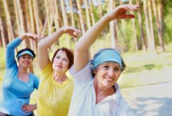 A recent study has asked whether there are links between a person being physically active or being sedentary can be associated with a higher risk of death. The question became – does the risk change at all if a person is genetically predisposed to live a longer life? Earlier research has indicated that low physical activity and more time spent sitting are linked with a higher risk of death.
  A new study has developed a novel method for studying age related disorders of the brain. The team has focused on the neurodegenerative disorder Huntington’s Disease for the research.