
Breathe Through Your Nose Not Your Mouth For Optimal Health
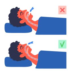 Our breath is the life source that keeps us going, and dodging any health issues related to it should be a top priority. As such, nasal breathing has increasingly gained attention as an essential element for respiratory well-being. By efficiently channeling air through your nose rather than your mouth, you are reaping numerous benefits for both short- and long-term health, whereas chronic mouth breathers may unknowingly be harmful in various ways.
By breathing through the nose, our bodies take up more oxygen due to an increase in airway resistance. This allows for improved elasticity and volume of the lungs, as well as increased oxygenation inside the nasal passageway thanks to a network of arteries, veins, capillaries and lymphatic vessels. A 2015 clinical review published by Nursing in General Practice found that these benefits can have a net result 10-20 percent higher uptake rate than when exhaling solely with your mouth.
Through the simple action of nasal breathing, you can promote a calming effect on both mind and body. This is because slow deep breaths stimulate your vagus nerve, which in turn activates your parasympathetic nervous system’s relaxation response. An additional bonus comes from nitric oxide production. This compound produced by nearly every cell helps relax blood vessels and boosts oxygen absorption capacity, while providing anti-fungal, antibacterial benefits.
Nasal breathing offers numerous benefits for your health and well being. Evidence indicates it can help keep the air moist, trap airborne debris before entering the body, reduce symptoms of common illnesses, encourage diaphragm action and even influence dental development such as creating arches in teeth.
Breathing through the mouth, which is done by a large portion of people, may have serious health implications such as gum disease or even sleep apnea. Additionally, it can also lead to dental decay and bad breath while influencing speech patterns and swallowing abilities.
Nasal breathing has been widely endorsed by many experts and cultures from around the world as a beneficial habit. While not everyone can easily breathe through their nose due to various issues such as a deviated septum or facial trauma, those who are able should cultivate this practice for its ample health perks.
Looking to become an expert breather? Try incorporating the following approaches into your daily life. Set reminders throughout the day, take a few minutes for breathing exercises like alternate nostril or diaphragmatic breath work, and consider taping your mouth at night. However, it’s important that this should not be attempted if any medical conditions apply like difficulty in nasal respiration or allergies/colds etc. One of the best mouth tapes is SomniFx, which can be purchased from Amazon and is easily applied and removed. Incorporating even just one of these strategies can help establish healthier habits as we move through our day-to-day lives.
To view the original scientific study click below:
The health benefits of nose breathing
 With Alzheimer’s and other forms of cognitive decline on the rise, could a lack of certain immune cells be to blame? Scientists are investigating whether replenishing these vital components could potentially help reverse some of this damage.
 With the prevalence of fast food and junk food on our menu, it’s no surprise that a Western-style diet high in fat and sugar can wreak havoc with human health. By consuming this type of diet it can cause obesity or diseases such as metabolic syndrome or diabetes. But how exactly does this type of eating affect our body internally?
  Research has revealed a potential link between water consumption and long-term health benefits. Staying hydrated could positively impact your overall well being. It has the potential to slow down the aging process by mitigating decreases in body water content which can result in higher levels of serum sodium.
 Tired of sitting hunched over your desk all day? Taking a five-minute stroll each half hour could be the secret to better health and improved mood, according to new research from Columbia University. Studies confirm that this simple movement can reduce blood sugar levels and spikes by up to 60%, while lowering your blood pressure and boosting your mood.
  A recent study has revealed a promising connection between Vitamin D levels in the human brain and lower rates of dementia. It suggests that higher amounts may be associated with decreased risk as people age, but further investigation is needed to fully comprehend how this crucial vitamin can influence cognition. While some possible causes for Alzheimer’s Disease are known, much more research into its origin remains an important area of inquiry.
 If you’re having trouble sleeping, now may be the time to take action. A recent Canadian study suggests a correlation between insomnia and an increased chance of developing memory decline or dementia as we age. Postdoc Nathan Cross details these findings in his longitudinal research which also highlights psychological disorders as potential comorbidities for diminished mental health.
 Paving the way for potential dietary treatments, researchers have revealed a promising link between our genes and what we eat. Exploring this connection could aid in treating serious conditions like diabetes, heart disease, and cancer. Recent research has sparked new insights into the potential benefits of intermittent fasting. According to a study published in Cell Metabolism, when mice only ate during certain periods of time, there were profound changes in gene expression impacting almost 80% of all genes. This can lead to improved blood sugar regulation and decreased risk for obesity as well as reversing signs typically associated with aging.
 Researchers at Iowa State have uncovered a groundbreaking discovery that the more body fat and less muscle mass we accumulate as we age, the lower our mental agility may become. An aging adult’s physical health can directly impact their cognitive abilities. This exciting development offers new hope for treatments that could help preserve an agile mind in older people who live a sedentary lifestyle or are obese due to natural changes with age.
 Some of us put on weight far more easily than others, and this may be related to our unique gut microbial compositions. Understanding how these microscopic organisms impact our health could help provide an explanation why some people seem unaffected by indulgent eating habits. A team at the Copenhagen University has uncovered a connection between weight gain and gut microbe composition. It appears some individuals may have an advantage in getting greater energy from food than those with less efficient microbes. This breakthrough research could explain why people sometimes struggle to achieve similar results despite eating identical meals.